Review of Radiographic Anatomy and Pathology
1

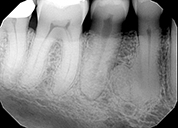
Radiograph 1. This patient has no symptoms. Describe the radiographic findings and provide a differential diagnosis.

2

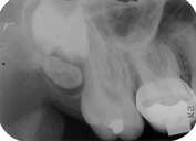
Radiograph 2. This patient presented for routine radiographs. Describe what your radiographic findings are in the upper right molar area.

3

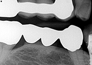
Radiograph 3. This mandibular bridge was placed about 10 years ago. The patient is asymptomatic. Describe your radiographic observations.

1

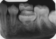
Radiograph 4. This 11 year old patient presented with a CC of a baby tooth which will not loosen. A radiograph was taken. Describe your radiographic findings.

2

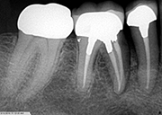
Radiograph 5. This patient has pain with endodonticaly treated #30. Describe the radiographic findings.

3

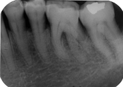
Radiograph 6. This patient presented to the periodontal department for scaling and root planing. What problems might be encounter with the debridement of #19?

2

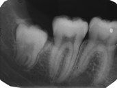
Radiograph 7. This 16 year old patient present evaluation of a third molar. Describe your radiographic findings for #32; is this tooth ready for extraction, what obstacles may be encountered?

3

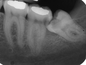
Radiograph 8. This patient is complaining of food impaction around the second molar. Why is this happening and what other problems do you see?