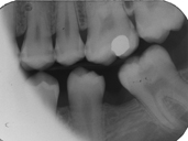
This patient presented for an initial examination and bitewing radiographs were taken. Describe what you see in this radiograph.
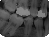
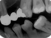
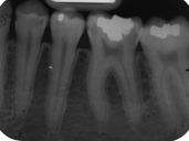
This patient presented for operative dentistry. Which teeth have caries and which surfaces are affected? Which molars are present in the MX and MN?
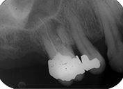
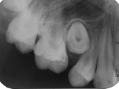
This patient presented with pain in the upper right molar area. A fistula was present and exudate was expressed. A radiograph was taken, however gutta percha was not placed in the sinus track. Describe what you see in this radiograph.
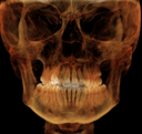
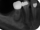
This patient presented for an initial examination. Describe what you see in this radiograph.
This patient presented for an initial examination and bitewing radiographs were taken. Describe what you see in this radiograph.
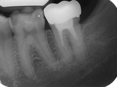
This patient presented for an initial examination and FMX radiographs were taken. Describe what you see in this radiograph.
This patient presented for an initial examination and FMX radiographs were taken. Describe what you see in this radiograph.
This patient presented for an initial examination and FMX radiographs were taken. Describe what you see in this radiograph.