logo
features Panoramic Anatomy

Maxillofacial anatomy on a panoramic radiograph is difficult. Variation in positioning acts to this complexity. Several structures are superimposed on this radiograph. In addition, the ghost images create another layer of complexity. Since panoramic radiography is a frequent imaging technique in the dental office, every dental student must exert sufficient efforts to understand the anatomy.

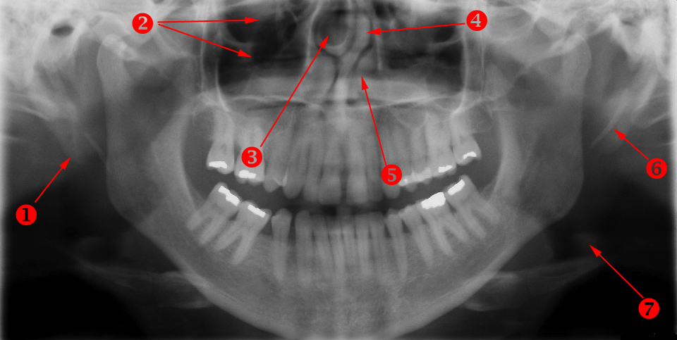
Panoramic Anatomy Case 4

Landmark 1: Right ear lobe

Landmark 2: (Bean shaped radiolucency) Anterior ethmoid air cell (Haller's cell)

Landmark 3: Concha bullosa

Landmark 4: Deviated nasal septum

Landmark 5: Inferior turbinate

Landmark 6: Left stylohyoid ligament

Landmark 7: Epiglottis