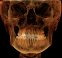
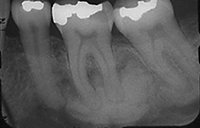
This patient presented for an initial examination and FMX radiographs were taken. Describe what you see in this radiograph.
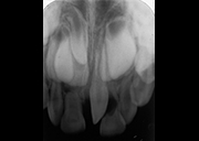
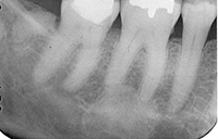
This patient presented for an initial examination and FMX radiographs were taken. Describe what you see in this radiograph.
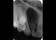
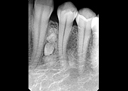
This patient presented for an initial examination and FMX radiographs were taken. Describe what you see in this radiograph.
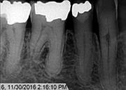
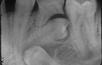
This middle aged female patient is asymptomatic. Please describe the radiographic findings and discuss plan for future treatment.
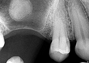
This patient presented for an initial examination and FMX radiographs were taken. Describe what you see in this radiograph.
This patient presented for an initial examination and FMX radiographs were taken. Describe what you see in this radiograph.
This patient presented for an initial examination and FMX radiographs were taken. Describe what you see in this radiograph.
This patient presented for an initial examination and FMX radiographs were taken. Describe what you see in this radiograph.