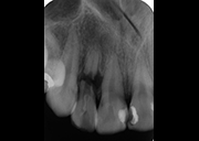
This patient presented for an initial examination and FMX radiographs were taken. Describe what you see in this radiograph.
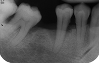
This patient presented for an initial examination and FMX radiographs were taken. Describe what you see in this radiograph.
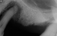
This patient presented for an initial examination and FMX radiographs were taken. Describe what you see in this radiograph.
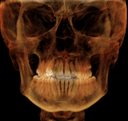
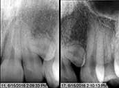
This patient presented for an initial examination. Describe what you see in this radiograph.Why did tooth #7 fracture?
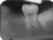
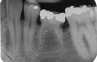
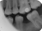
This patient presented for an initial examination and bitewing radiographs were taken. Describe what you see in this radiograph.
This patient presented for an initial examination and FMX radiographs were taken. Describe what you see in this radiograph.
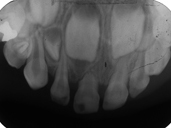
Identify the relationship of the supernumerary tooth with the lateral incisor and the canine. Other than the position, what additinal information is needed for successful manangement of this patient?
This patient presented for an initial examination and FMX radiographs were taken. Describe what you see in this radiograph.