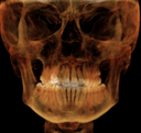
This patient has no symptoms. Describe the radiographic findings and provide a differential diagnosis.
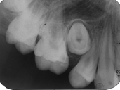
This patient presented for routine radiographs. Describe what your radiographic findings are in the upper right molar area.
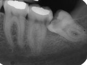
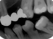
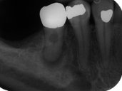
This patient is complaining of food impaction around the second molar. Why is this happening and what other problems do you see?
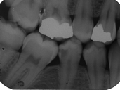
This patient presented for operative dentistry. Which teeth have caries and which surfaces are affected? Which molars are present in the MX and MN?
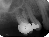
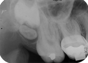
This patient presented with pain in the upper right molar area. A fistula was present and exudate was expressed. During radiography gutta percha was not placed in the sinus track. Describe what you see in this radiograph.
This patient presented for an initial examination. Describe what you see in this radiograph. What are your clinical concerns? What additional images would you acquire to address your concern?
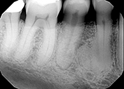
This patient presented for an initial examination and bitewing radiographs were taken. Describe what you see in this radiograph.
This patient presented for an initial examination and FMX radiographs were taken. Describe what you see in this radiograph.