logo
features Intraoral Radiography

During each intraoral session, we will attempt to interpret 12-13 intraoral images. Each session will end with one intraoral image for competency. Course requirement is interpreting 25 intraoral images and successfully completing two competency images. For each image, please identify the radiograph (e.g. maxillary right molar periapical film), evaluate any technical discrepancy, and then describe the major findings. Your teacher will quiz you on anatomic landmarks.

Review of Radiographic Anatomy and Pathology
1

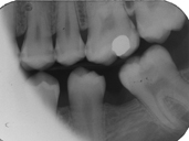
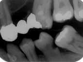
Radiograph 9. This patient presented for an initial examination and bitewing radiographs were taken. Describe what you see in this radiograph.

2

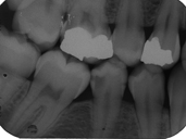
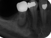
Radiograph 10. This patient presented for operative dentistry. Which teeth have caries and which surfaces are affected? Which molars are present in the MX and MN?

3

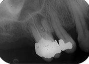
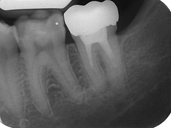
Radiograph 11. This patient presented with pain in the upper right molar area. A fistula was present and exudate was expressed. A radiograph was taken, however gutta percha was not placed in the sinus track. Describe what you see in this radiograph.

1

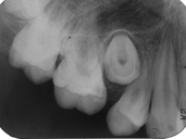
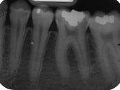
Radiograph 12. This patient presented for an initial examination. Describe what you see in this radiograph.

2

Radiograph 13. This patient presented for an initial examination and bitewing radiographs were taken. Describe what you see in this radiograph.

3

Radiograph 14. This patient presented for an initial examination and FMX radiographs were taken. Describe what you see in this radiograph.

2

Radiograph 15. This patient presented for an initial examination and FMX radiographs were taken. Describe what you see in this radiograph.

3

Radiograph 16. This patient presented for an initial examination and FMX radiographs were taken. Describe what you see in this radiograph.